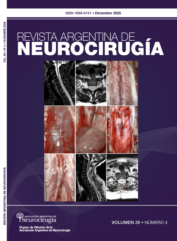
Anatomy of the corticospinal tract and its importance in the resection of pontine cavernomas
DOI:
https://doi.org/10.59156/revista.v39i04.787Keywords:
Corticospinal, Dissection, Pontine cavernoma, TractographyAbstract
Background: surgical resection of pontine cavernomas presents a challenge due to the density of critical neural structures in this region, including the corticospinal tract.
Objectives: to provide a detailed description of the corticospinal tract's course using dissections and tractography, in order to define anatomical parameters that allow for the avoidance of injury to this tract during pontine surgery.
Methods: tractography was performed using DSI Studio software on 1065 brains from healthy subjects belonging to the Human Connectome Project to illustrate the corticospinal tract. Six brainstems were designed using microsurgical instruments and the Klingler method.
Results: in the tractography, the TCE fibers were found ventrally related to the superficial layer of the ESTP fibers and dorsally to the EPTP, separated from the pontine tegmentum by the LM. Relationships of the TCE in dissections: ventrally, 1.1-2 mm of the ESTP was resected. Laterally, the EMTP was identified, and a resection was performed to the apparent origin of CN V, located 3.9-4.1 mm from the TCE. Deep dissection allowed the spinal trigeminal nucleus to be located 10.2-12.4 mm deep. Dorsally, the EPTP was identified, separating the pyramidal fibers from the LM.
Conclusion: the results obtained allow for the definition of specific anatomical parameters as key surgical landmarks. The arrangement of transverse fibers can be used during the planning and execution of pontine surgical approaches.
Downloads
References
1. Cervio A, Giovanini S, Nuñez M, Caffaratti G, Grijalba Romero M, Villamil F, y col. Tratamiento quirúrgico de los cavernomas de tronco cerebral. Rev Latinoam Neurocir. 2020;29(5):90-119.
2. Catapano JS, Rumalla K, Srinivasan VM, Lawrence PM, Keil KL, Lawton MT. A taxonomy for brainstem cavernous malformations: subtypes of medullary lesions. J Neurosurg. 2022;138(1):128-46. DOI: https://doi.org/10.3171/2022.3.JNS22626
3. Catapano JS, Rumalla K, Srinivasan VM, Lawrence PM, Keil KL, Lawton MT. A taxonomy for brainstem cavernous malformations: subtypes of midbrain lesions. J Neurosurg. 2021;136(6):1667-86. DOI: https://doi.org/10.3171/2021.8.JNS211694
4. Cantore G, Missori P, Santoro A. Cavernous angiomas of the brain stem. Surg Neurol. 1999;52:84-94. DOI: https://doi.org/10.1016/S0090-3019(99)00036-1
5. Bello L, Castellano A, Fava E, Casaceli G, Riva M, Scotti G, et al. Intraoperative use of diffusion tensor imaging fiber tractography and subcortical mapping for resection of gliomas: technical considerations. Neurosurg Focus. 2010;28(2):E6. DOI: https://doi.org/10.3171/2009.12.FOCUS09240
6. Fernández-Miranda JC, Pathak S, Engh J, Jarbo K, Verstynen T, Yeh FC, y col. High-definition fiber tractography of the human brain: neuroanatomical validation and neurosurgical applications. Neurosurgery. 2012;71(2):430-53. DOI: https://doi.org/10.1227/NEU.0b013e3182592faa
7. Maesawa S, Fujii M, Nakahara N, Watanabe T, Wakabayashi T, Yoshida J. Intraoperative tractography and motor evoked potential (MEP) monitoring in surgery for gliomas around the corticospinal tract. World Neurosurg. 2010;74(1):153-61. DOI: https://doi.org/10.1016/j.wneu.2010.03.022
8. Yagmurlu K, Rhoton AL Jr, Tanriover N, Bennett JA. Three-dimensional microsurgical anatomy and the safe entry zones of the brainstem. Oper Neurosurg (Hagerstown). 2014;10(4):602-20. DOI: https://doi.org/10.1227/NEU.0000000000000466
9. Berman JI, Berger MS, Mukherjee P, Henry RG. Diffusion-tensor imaging-guided tracking of fibers of the pyramidal tract combined with intraoperative cortical stimulation mapping in patients with gliomas. J Neurosurg. 2004;101(1):66-72. DOI: https://doi.org/10.3171/jns.2004.101.1.0066
10. Ordóñez-Rubiano EG. Anatomía microquirúrgica en 3D del tracto corticoespinal y de la vía del lemnisco basada en microdisección de fibras y demostración a través de tractografía. Neurocirugía. 2019;30(6):309-10. DOI: https://doi.org/10.1016/j.neucir.2018.11.005
11. Kovanlikaya I, Firat Z, Kovanlikaya A, Uluğ AM, Cihangiroglu MM, John M, y col. Assessment of the corticospinal tract alterations before and after resection of brainstem lesions using Diffusion Tensor Imaging (DTI) and tractography at 3T. Eur J Radiol. 2011;77(3):383-91. DOI: https://doi.org/10.1016/j.ejrad.2009.08.012
12. Chenot Q, Tzourio-Mazoyer N, Rheault F, Descoteaux M, Crivello F, Zago L, y col. A population-based atlas of the human pyramidal tract in 410 healthy participants. Brain Struct Funct. 2019;224(2):599-612. DOI: https://doi.org/10.1007/s00429-018-1798-7
13. Recalde RJ, Figueiredo EG, de Oliveira E. Microsurgical anatomy of the safe entry zones on the anterolateral brainstem related to surgical approaches to cavernous malformations. Oper Neurosurg (Hagerstown). 2008;62(Suppl 3):9-17. DOI: https://doi.org/10.1227/01.neu.0000317368.69523.40
14. Yao Y, Ulrich NH, Guggenberger R, Alzarhani YA, Bertalanffy H, Kollias SS. Quantification of corticospinal tracts with diffusion tensor imaging in brainstem surgery: prognostic value in 14 consecutive 159 cases at 3T magnetic resonance imaging. World Neurosurg. 2015;83(6):1006-14. DOI: https://doi.org/10.1016/j.wneu.2015.01.045
15. Kwon HG, Son SM, Chang MC, Kim S, Kwon YH, Jang SH. Characteristics of the aberrant pyramidal tract in comparison with the pyramidal tract in the human brain. BMC Neurosci. 2011;12:108. DOI: https://doi.org/10.1186/1471-2202-12-108
16. Benner D, Hendricks BK, Benet A, Graffeo CS, Scherschinski L, Srinivasan VM, y col. A system of anatomical triangles defining dissection routes to brainstem cavernous malformations: definitions and application to a cohort of 183 patients. J Neurosurg. 2022;138(3):768-84. DOI: https://doi.org/10.3171/2022.6.JNS212907
17. Abla AA, Lekovic GP, Turner JD, de Oliveira JG, Porter R, Spetzler RF. Advances in the treatment and outcome of brainstem cavernous malformation surgery: a single-center case series of 300 surgically treated patients. Neurosurgery. 2011;68(2):403-14. DOI: https://doi.org/10.1227/NEU.0b013e3181ff9cde
18. Vassal F, Schneider F, Nuti C. Intraoperative use of diffusion tensor imaging-based tractography for resection of gliomas located near the pyramidal tract: comparison with subcortical stimulation mapping and contribution to surgical outcomes. Br J Neurosurg. 2013;27(5):668-75. DOI: https://doi.org/10.3109/02688697.2013.771730
19. Catapano JS, Rumalla K, Srinivasan VM, Lawrence PM, Keil KL, Lawton MT. A taxonomy for brainstem cavernous malformations: subtypes of pontine lesions. Part 1: basilar, peritrigeminal, and middle peduncular. J Neurosurg. 2022;137(5):1462-76. DOI: https://doi.org/10.3171/2022.1.JNS212690