WISTAR RATS AS A MODEL OF MICRO SURGICAL EDUCATION: ANATOMY AND CLASSICAL EXERCISES
DOI:
https://doi.org/10.59156/revista.v36i1.275Keywords:
Wistar rats, anatomy, vascular anastomosis, microsurgical training.Abstract
ABSTRACT:
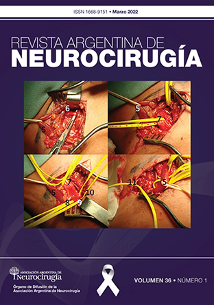
Introduction: The use of rodents as live models for microvascular training constitutes one of the most widespread practical methods. The objective of the present work is to describe the microsurgical anatomy of the Wistar rat in the cervical and femoral access routes, for the training of classic vascular anastomosis exercises.
Materials and methods: The procedures were carried out in the Central Animal Laboratory of the Faculty of Pharmacy and Biochemistry of the University of Buenos Aires. Two rats of the Wistar species were used in which end-to-end and end-to-side anastomoses of the cervical and femoral region were performed respectively.
Results: anatomy of the cervical and femoral region, made up of muscle groups, fasciae, glands and neurovascular bundles. A cervical approach was made with the end-to-end anastomosis of the Common Carotid Artery. In the other specimen, a left femoral access and end-to-end anastomosis were made between the Femoral Vein and the Superficial Caudal Epigastric Vein. Adequate vascular permeability was verified by Doppler ultrasound.Conclusion: The use of live models for microvascular training is extremely necessary and effective to acquire microsurgical skills in our training as neurosurgeons. Being a classic simulation and training model, it is essential to acquire the anatomical knowledge of the specimens, as well as the exercise of vascular anastomosis techniques.
Downloads